
Thymosin Alpha-1: Mechanism of Action
A mechanism-focused reading of Thymosin Alpha-1: from its acetylated 28-residue sequence and NMR-resolved conformation to the divergent TLR9 (pDC) and TLR2 (mDC) signaling arms, IFNAR/IDO tryptophan catabolism, and the structural questions still open in the literature. Educational reference.

For research use only. Not for human consumption. This article is educational reference material. It is not medical advice and is not a recommendation to use any substance.
From sequence to signal: why the mechanism is unusual
Thymosin Alpha-1 (Tα1) is an acetylated 28-residue peptide, and its reported pharmacology does not map neatly onto the single-receptor, single-pathway picture common to many signaling peptides. Instead, the peer-reviewed literature describes a mechanism defined by divergence: the same peptide is reported to engage different Toll-like receptors (TLRs) in different dendritic cell subsets, producing opposing immunological outputs from what appears to be one molecular input.
This article reads the mechanism from the molecule outward, starting with what the sequence and solution structure permit, moving through the two divergent TLR arms characterized by Romani and colleagues at the University of Perugia, and ending with the structural questions the field has not yet answered. A broader treatment of the compound's chemistry, classification, and regulatory status is available in the Thymosin Alpha-1 research overview, and the primary study record is summarized in the Thymosin Alpha-1 published research article.
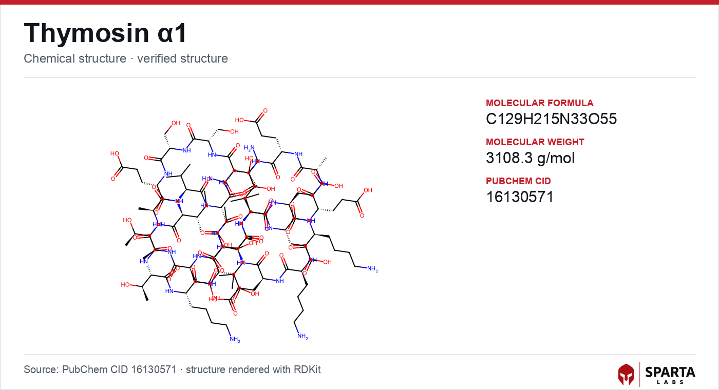
Figure: chemical structure of Thymosin Alpha-1.
What the molecule offers a receptor
Any mechanistic account is constrained by the physical peptide. The 2012 NMR characterization by Nepravishta and colleagues in FEBS J described the solution-state conformation of Tα1 as a distorted N-terminal helical region paired with a defined C-terminal alpha-helix spanning approximately residues 14 to 26 [1]. Findings from research models do not establish safety or efficacy in humans. Sparta Labs makes no claims about the use of this compound.
That structural description matters for a mechanistic reading for two reasons. First, a partially ordered peptide with a discrete helical face presents a candidate interaction surface, but the NMR work stopped short of describing any receptor contact. Second, the conformation was resolved in isolation rather than in complex, so it constrains hypotheses about how Tα1 might dock without confirming any of them. The authors framed the structure as a foundation for subsequent structural pharmacology rather than as a completed binding model.
The plasmacytoid arm: TLR9 / MyD88 / IRF7
The first of the two divergent arms was characterized in the antiviral context. The 2007 study by Romani and colleagues in International Immunology reported that Tα1 activated a TLR9/MyD88/IRF7-dependent pathway in plasmacytoid dendritic cells (pDCs) during murine cytomegalovirus (mCMV) infection in vivo [2].
The causal weight of the finding came from genetics rather than correlation. The authors used TLR9-knockout mice to show that the antiviral response observed in wild-type animals was substantially abrogated when TLR9 signaling was genetically disrupted, indicating that TLR9 was required in the cascade rather than merely coincident with it [2]. Downstream, MyD88-dependent signaling was reported to activate interferon regulatory factor 7 (IRF7), the transcription factor that drives type I interferon gene expression, linking receptor engagement to IFN-α output through a defined intermediate.
The myeloid arm: TLR2 and a different output
The second arm diverges at the level of both receptor and cytokine. Within the same 2007 study, myeloid dendritic cells (mDCs) were reported to use TLR2 rather than TLR9 in response to Tα1, and to produce IL-12 p70 rather than the interferon-dominated profile of the pDC arm [2]. The same peptide, in other words, was associated with different receptor utilization and different signaling endpoints depending on the responding cell type.
A 2016 review by Shrivastava and John in Expert Opinion on Biological Therapy synthesized these observations into a parallel-arms model: pDCs responding via TLR9 were associated with IFN-α and IL-10 outputs, while mDCs responding via TLR2 were associated with IL-12 p70 [3]. This cell-type-specific receptor split is a defining feature of the reported Tα1 mechanism, and it distinguishes the peptide's characterized pharmacology from single-receptor signaling molecules. A structurally unrelated innate-immune mechanism, centered on MC1R and NF-κB rather than TLR signaling, is described for the copper-binding tripeptide in the GHK-Cu mechanism of action article, and a melanocortin-pathway contrast is covered in the KPV mechanism of action article.
Converging on tryptophan catabolism: the IFNAR / IDO axis
The two arms do not stay wholly separate. The earlier 2006 Blood study by Romani and colleagues described a downstream node where TLR9 signaling fed into a second-messenger loop [4]. In that model, Tα1 signaling via TLR9 drove type I interferon production, which then activated the type I interferon receptor (IFNAR) on the same or neighboring dendritic cells.
IFNAR signaling was identified as required for expression of indoleamine 2,3-dioxygenase (IDO), the rate-limiting enzyme of tryptophan catabolism along the kynurenine pathway [4]. Local tryptophan depletion by IDO was, in turn, associated with differentiation of CD4+ regulatory T cells (Tregs) in the experimental models studied. The authors described this as a mechanism by which Tα1 could concurrently drive innate antiviral and antifungal responses through one arm while establishing tolerance-favoring conditions through IDO, a duality the paper summarized as "a regulatory environment for balance of inflammation and tolerance."
This dual capacity is the mechanistic through-line that connects the divergent receptor arms: interferon-driven activation and IDO-driven regulation are reported as outputs of a single upstream trigger rather than as separate phenomena.
Older observations the TLR model helped contextualize
Not all Tα1 mechanistic observations began with the TLR framework. A 2007 review by Goldstein in Ann N Y Acad Sci, surveying the broader thymosin research history, noted that earlier in vitro studies associated Tα1 with upregulation of major histocompatibility complex (MHC) class I molecule expression on cell surfaces [5]. That observation predates the receptor-pathway work but is consistent with the downstream IFN-γ signaling described in the later TLR9-pathway studies, and the mechanistic work provided a plausible upstream anchor for what had previously been an isolated finding.
The reported cytokine profile is similarly broad. A 2020 comprehensive review by Dominari and colleagues in World J Virol summarized IL-12, IL-10, IFN-α, and IFN-γ as frequently cited outputs across dendritic-cell-focused experimental systems, while noting that the direction and magnitude of these associations varied across cell types, disease models, and study conditions [6]. That context-dependence is itself a mechanistic characteristic worth noting for researchers designing cell-type- or model-specific investigations, and it aligns with the parallel-arms structure described above.
Limits of current understanding
Several boundaries constrain what the mechanism can currently be said to establish.
The dominant TLR9/pDC framework is derived primarily from murine systems. A sepsis study by Han and colleagues in Crit Care examined TLR2, TLR4, and MyD88 mRNA expression on human peripheral blood mononuclear cells during Tα1 treatment, providing preliminary in-human signal data consistent with the preclinical TLR-pathway findings [7]. That report indicates the murine-to-human translation is an active line of inquiry rather than an untested assumption, but it does not substitute for higher-resolution human mechanistic work.
The structural basis of Tα1's interaction with TLR9 or any co-receptor complex has not been described in a ligand-receptor co-crystal structure. Combined with the isolated-peptide NMR conformation [1], this leaves the fundamental question open: whether Tα1 acts as a direct TLR9 ligand, an indirect modulator through accessory molecules, or through a related mechanism has not been resolved in the literature. Subsequent work from the same Perugia group, cited in a 2007 review by Romani and colleagues in Ann N Y Acad Sci, extended the dual-arm findings into murine hematopoietic stem cell transplantation models where antifungal activity and alloantigen tolerance were investigated together [8], but that extension operated within, rather than beyond, the existing mechanistic frame.
Research-grade Thymosin Alpha-1 from Sparta Labs is supplied for laboratory research use and supported by third-party analytical verification.
References
-
Nepravishta R, Mandaliti W, Valente S, Alagna NS, Labruna S, Pica F, et al. NMR structure of human thymosin alpha-1. FEBS J. 2012;279(3):401–409. PMID: 22115779. DOI: 10.1111/j.1742-4658.2011.08428.x. View on PubMed
-
Romani L, Bistoni F, Montagnoli C, Gaziano R, Bozza S, Fallarino F, et al. Thymosin alpha1 activates the TLR9/MyD88/IRF7-dependent murine cytomegalovirus sensing for induction of anti-viral responses in vivo. Int Immunol. 2007;19(10):1261–1271. PMID: 17804687. DOI: 10.1093/intimm/dxm099. View on PubMed
-
Shrivastava R, John SM. Immune modulation with thymosin alpha 1 treatment. Expert Opin Biol Ther. 2016;16(9):1147–1153. PMID: 27450734. DOI: 10.1080/14712598.2016.1198809. View on PubMed
-
Romani L, Bistoni F, Gaziano R, Bozza S, Montagnoli C, Perruccio K, et al. Thymosin alpha 1 activates dendritic cell tryptophan catabolism and establishes a regulatory environment for balance of inflammation and tolerance. Blood. 2006;108(7):2265–2274. PMID: 16741252. DOI: 10.1182/blood-2006-02-004762. View on PubMed
-
Goldstein AL. History of the discovery of the thymosins. Ann N Y Acad Sci. 2007;1112:1–13. PMID: 17600284. DOI: 10.1196/annals.1415.001. View on PubMed
-
Dominari A, Hathaway D 3rd, Pandav K, Vasan S, Dhindsa DS, Dave K, et al. Thymosin alpha 1: A comprehensive review of the literature. World J Virol. 2020;9(5):67–78. PMID: 33362999. PMC7747025. DOI: 10.5501/wjv.v9.i5.67. Full text on PMC
-
Han S, Sun H, He F, Zheng H. Changes of TLR2, TLR4, MyD88 mRNA expressions on peripheral blood mononuclear cells in severe sepsis patients during treatment with thymosin α1. Crit Care. 2015;19(Suppl 1):P15. PMC4796653. Source: ncbi.nlm.nih.gov
-
Romani L, Montagnoli C, Bozza S, Perruccio K, Spreca A, Allavena P, et al. Thymosin alpha1 overview: an approach towards immune reconstitution. Ann N Y Acad Sci. 2007;1112:326–338. PMID: 17495242. DOI: 10.1196/annals.1415.006. View on PubMed
Disclaimer. Statements in this article have not been evaluated by the Food and Drug Administration. This compound is not intended to diagnose, treat, cure, or prevent any disease. Sparta Labs sells research-use-only materials. Content is provided for educational and informational purposes only and does not constitute medical advice. Consult a qualified medical professional for any health concerns.
Frequently asked questions
Is Thymosin Alpha-1 believed to be a direct TLR9 ligand?
The published literature has not resolved this. No ligand-receptor co-crystal structure of Thymosin Alpha-1 with TLR9 or an accessory complex has been reported. Romani and colleagues demonstrated that TLR9 signaling was required for the observed responses using knockout models, but whether the peptide binds TLR9 directly, acts through accessory molecules, or engages a related mechanism remains an open research question.
Why does Thymosin Alpha-1 signal through two different Toll-like receptors?
Published murine studies report a cell-type split: plasmacytoid dendritic cells appear to respond via the TLR9/MyD88/IRF7 axis, while myeloid dendritic cells appear to use TLR2. Shrivastava and colleagues characterized these as parallel arms, with the pDC route associated with type I interferon and IL-10 outputs and the mDC route associated with IL-12 p70. The two-receptor split is a defining feature of the reported pharmacology rather than an inconsistency.
What is the IDO pathway and how does it appear in Thymosin Alpha-1 research?
Indoleamine 2,3-dioxygenase (IDO) is the rate-limiting enzyme of tryptophan catabolism along the kynurenine pathway. Romani and colleagues reported in Blood (2006) that Thymosin Alpha-1 signaling through TLR9 drove type I interferon production, which activated IFNAR signaling required for subsequent IDO expression in dendritic cells, an event associated with regulatory T cell generation in the models studied.
What does the NMR structure of Thymosin Alpha-1 describe?
The 2012 NMR characterization by Nepravishta and colleagues described the solution-state conformation of the 28-residue peptide as a distorted N-terminal helical region with a defined C-terminal alpha-helix spanning approximately residues 14 to 26. This conformational description is cited as a foundation for structural pharmacology work, though it does not by itself establish how the peptide engages any receptor.
Has the Thymosin Alpha-1 mechanism been confirmed in human cells?
The dominant TLR9/pDC framework is derived largely from murine systems. A sepsis study by Han and colleagues examined TLR2, TLR4, and MyD88 mRNA expression on human peripheral blood mononuclear cells during Thymosin Alpha-1 treatment, providing preliminary in-human signal data consistent with preclinical findings. Higher-resolution human mechanistic studies remain an active area of investigation.