
KPV Mechanism of Action: Published Research
A mechanism-focused review of KPV (Lys-Pro-Val), the C-terminal tripeptide of α-MSH: its receptor-independent action on importin-α and NF-κB nuclear import, structural mimicry of the IL-1β C-terminus, and PepT1-mediated cellular uptake as documented in primary literature.

For research use only. Not for human consumption. This article is educational reference material. It is not medical advice and is not a recommendation to use any substance.
Introduction
KPV is a tripeptide with the sequence lysine-proline-valine (Lys-Pro-Val), corresponding to residues 11 through 13 of alpha-melanocyte-stimulating hormone (α-MSH). Because it represents only the carboxy-terminal fragment of a much larger signaling peptide, KPV has been studied as a way to isolate the anti-inflammatory character of the melanocortin system from the pigmentary and other receptor-mediated activities of the full-length hormone. The mechanistic literature is notable because the most frequently reported mode of action is receptor-independent: rather than engaging a cell-surface melanocortin receptor, KPV has been reported to act inside the cell on the nuclear-transport machinery that carries the transcription factor NF-κB into the nucleus. This article summarizes the reported molecular interactions and the experimental systems in which they were observed, with attribution to the primary literature.
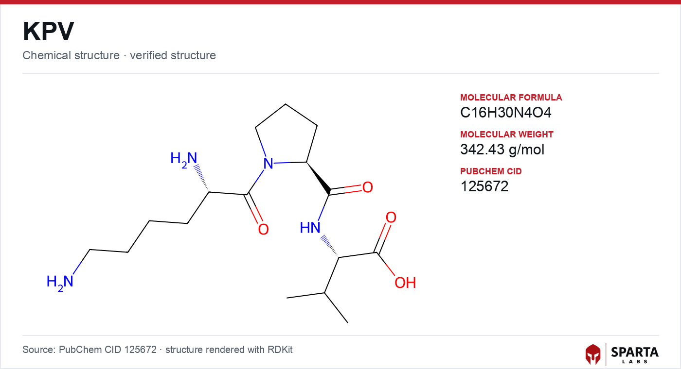
Figure: chemical structure of KPV (Lys-Pro-Val).
From a hormone fragment to a standalone probe
α-MSH is a 13-residue peptide processed from pro-opiomelanocortin. Its melanocortin activity is concentrated in the core sequence, while immunomodulatory activity has been mapped, in part, to the C-terminal tripeptide. Getting, Schiöth, and Perretti dissected these contributions in a 2003 study in the Journal of Pharmacology and Experimental Therapeutics, comparing the core pharmacophore against the C-terminal KPV sequence in a murine peritonitis model [1]. They reported that KPV produced anti-inflammatory effects through a route distinct from the core fragment and concluded that KPV was "unlikely to mediate its effects through melanocortin receptors," pointing instead toward interference with interleukin-1β (IL-1β) function [1].
Findings from research models do not establish safety or efficacy in humans. Sparta Labs makes no claims about the use of this compound.
This receptor-independent framing was reinforced by Luger and Brzoska, who noted in a 2007 review in the Annals of the Rheumatic Diseases that the tripeptide "seems not to bind to MC-1R and fails to increase cyclic adenosine monophosphate (cAMP) levels" [2]. That observation is what separates KPV mechanistically from full-length melanocortin agonists such as those discussed in the melanotan-2 mechanism of action and PT-141 mechanism of action literature, both of which depend on canonical melanocortin receptor engagement and cAMP signaling. For a broader treatment of KPV's classification and lineage, the KPV research overview provides additional context.
The importin-α model: blocking NF-κB at the nuclear gate
NF-κB is a transcription factor held inactive in the cytoplasm by its inhibitor IκBα. Upon inflammatory stimulation, IκBα is phosphorylated and degraded, freeing the p65RelA subunit to enter the nucleus. Nuclear entry depends on importin-α proteins, which recognize the nuclear localization signal (NLS) on p65RelA and shuttle it through the nuclear pore.
Land published a granular account of KPV's molecular action in 2012, using human bronchial epithelial cells stimulated with tumor necrosis factor-α (TNF-α) [3]. Employing co-immunoprecipitation and GST pull-down assays, the study reported that KPV translocated into the nucleus and reduced the binding of importin-α3 to p65RelA, thereby limiting nuclear import of the active NF-κB subunit. The study reported that this occurred without altering upstream IκBα phosphorylation, situating KPV's reported action downstream of the classical activation cascade and at the level of nuclear transport itself [3].
Computational docking in the same study placed KPV in the vicinity of armadillo repeat domains 7 and 8 of importin-α, regions recognized as important for NLS-cargo recognition [3]. By occupying or interfering with this recognition surface, KPV was proposed to impede the importin-α–p65RelA interaction. The study noted a parallel role for MC3R agonism in the same epithelial system, raising the possibility that a receptor-independent transport-blockade route and a receptor-mediated cAMP route could converge on overlapping inflammatory outputs [3]. Interference with NF-κB-dependent signaling has also been described for other repair-cluster peptides; the BPC-157 mechanism of action literature discusses a distinct set of pathways sometimes compared alongside KPV.
Structural mimicry of the IL-1β C-terminus
A second proposed interaction rests on sequence resemblance. The Lys-Pro-Val motif corresponds to the C-terminal tripeptide of IL-1β (residues 193 to 195), and this overlap has been offered as a structural rationale for KPV's reported effects on IL-1 signaling. Luger and Brzoska reported that KPV and its stereoisomeric derivative K(D)PT interfered with the surface binding of radiolabeled IL-1β to T cells, consistent with antagonism at the IL-1 receptor complex or an accessory binding site [2].
Getting and colleagues' peritonitis data provided in vivo support for IL-1β-directed modulation attributable to the C-terminal sequence, distinct from the macrophage-directed effects assigned to the α-MSH core [1]. Taken together, the two mechanistic threads — nuclear-import interference and IL-1β mimicry — are not mutually exclusive, since IL-1β is itself an upstream activator of NF-κB, and both would be expected to dampen the same downstream transcriptional program.
PepT1: a route to the intracellular compartment
An intracellular mechanism raises an obvious question: how does a hydrophilic tripeptide reach the cytoplasm and nucleus? Dalmasso and colleagues addressed this in a 2008 study in Gastroenterology, reporting that KPV is transported into intestinal epithelial cells by PepT1 (SLC15A1), the proton-coupled oligopeptide transporter expressed at the apical surface of small-intestinal epithelium [4]. Following uptake, KPV at nanomolar concentrations was reported to reduce NF-κB and MAP-kinase signaling and pro-inflammatory cytokine secretion in intestinal epithelial cell lines [4].
The PepT1 route is significant because it links a plausible delivery mechanism to the intracellular targets proposed by Land, and because it implies that the tissues where KPV is most active may partly reflect where PepT1 is expressed. The extent to which non-intestinal tissues support comparable PepT1-mediated entry remains an open question in the literature [4].
Cross-tissue observations
Reported effects of KPV span several experimental systems, which is part of why the receptor-independent, transporter-delivered model has attracted interest. In bronchial epithelium, Land reported reductions in NF-κB activity and in secreted IL-8, eotaxin, and matrix metalloproteinase-9 (MMP-9) activity following KPV application [3]. In murine models of intestinal inflammation, Kannengiesser and colleagues (2008) reported reduced pro-inflammatory cytokine expression and diminished mucosal inflammatory infiltrate on histology [5]. In a mouse traumatic-brain-injury model, Schaible and colleagues (2013) reported that the α-MSH(11–13) tripeptide sequence was associated with reduced secondary lesion volume, decreased neuronal apoptosis, and attenuated microglial activation relative to vehicle controls [6]. These observations sit within a broader body of work on melanocortin regulation of inflammation summarized by Wang and colleagues (2019) [7].
None of these findings establish a mechanism in humans, and the studies describe distinct model systems that are not directly interchangeable. They are presented here as reported observations rather than conclusions about clinical activity.
Limits of current understanding
Several elements of the KPV mechanism remain provisional. The importin-α docking geometry proposed by Land is computational rather than crystallographic; a co-complex structure at atomic resolution has not been established, and it defines a clear direction for future structural work [3]. The relative contributions of the importin-α route, the IL-1β-mimicry route, and any residual melanocortin-receptor involvement across different tissues are not fully resolved [1][3]. Finally, the generality of PepT1-mediated entry beyond intestinal epithelium is unsettled [4]. Researchers examining these pathways can reference the KPV published research summary for study-by-study detail, and material specifications are documented for KPV from Sparta Labs.
References
-
Getting SJ, Schiöth HB, Perretti M. Dissection of the anti-inflammatory effect of the core and C-terminal (KPV) alpha-melanocyte-stimulating hormone peptides. J Pharmacol Exp Ther. 2003;306(2):631-637. PMID: 12750433. DOI: 10.1124/jpet.103.051623
-
Luger TA, Brzoska T. alpha-MSH related peptides: a new class of anti-inflammatory and immunomodulating drugs. Ann Rheum Dis. 2007;66(Suppl 3):iii52-iii55. PMID: 17934097. DOI: 10.1136/ard.2007.079780
-
Land SC. Inhibition of cellular and systemic inflammation cues in human bronchial epithelial cells by melanocortin-related peptides: mechanism of KPV action and a role for MC3R agonists. Int J Physiol Pathophysiol Pharmacol. 2012;4(2):59-73. PMID: 22837805
-
Dalmasso G, Charrier-Hisamuddin L, Nguyen HTT, Yan Y, Sitaraman S, Merlin D. PepT1-mediated tripeptide KPV uptake reduces intestinal inflammation. Gastroenterology. 2008;134(1):166-178. PMID: 18061177. DOI: 10.1053/j.gastro.2007.10.026
-
Kannengiesser K, Maaser C, Heidemann J, Luegering A, Ross M, Brzoska T, Böhm M, Luger TA, Domschke W, Kucharzik T. Melanocortin-derived tripeptide KPV has anti-inflammatory potential in murine models of inflammatory bowel disease. Inflamm Bowel Dis. 2008;14(3):324-331. PMID: 18092346. DOI: 10.1002/ibd.20334
-
Schaible EV, Steinsträßer A, Jahn-Eimermacher A, Luh C, Sebastiani A, Kornes F, Pieter D, Schäfer MKE, Engelhard K, Thal SC. Single administration of tripeptide α-MSH(11–13) attenuates brain damage by reduced inflammation and apoptosis after experimental traumatic brain injury in mice. PLoS One. 2013;8(8):e71056. PMID: 23940690. DOI: 10.1371/journal.pone.0071056
-
Wang W, Guo DY, Lin YJ, Tao YX. Melanocortin regulation of inflammation. Front Endocrinol. 2019;10:683. PMID: 31649620. DOI: 10.3389/fendo.2019.00683
Disclaimer. Statements in this article have not been evaluated by the Food and Drug Administration. This compound is not intended to diagnose, treat, cure, or prevent any disease. Sparta Labs sells research-use-only materials. Content is provided for educational and informational purposes only and does not constitute medical advice. Consult a qualified medical professional for any health concerns.
Frequently asked questions
Is KPV a melanocortin receptor agonist?
Published research indicates that KPV does not behave like a classical melanocortin receptor agonist. Luger and Brzoska (2007) reported that the tripeptide does not appreciably bind MC1R or raise cAMP, and Getting and colleagues (2003) concluded its activity was unlikely to be mediated through melanocortin receptors. This distinguishes KPV mechanistically from full-length α-MSH analogs such as melanotan-2.
What is the significance of KPV resembling the IL-1β C-terminus?
The Lys-Pro-Val sequence corresponds to the C-terminal tripeptide of interleukin-1β (residues 193 to 195). Luger and Brzoska (2007) reported that KPV and its derivative interfered with IL-1β binding to T cells, and researchers have proposed this structural resemblance as one basis for KPV's reported interference with IL-1 signaling in experimental systems.
How is KPV proposed to reach intracellular targets?
Dalmasso and colleagues (2008), publishing in Gastroenterology, reported that KPV is transported into intestinal epithelial cells by PepT1 (SLC15A1), a proton-coupled oligopeptide transporter. This uptake route offers a mechanistic explanation for how a small tripeptide could reach intracellular signaling and nuclear-transport machinery at low concentrations.
What is the importin-α model of KPV action?
Land (2012) reported that in human bronchial epithelial cells KPV translocated to the nucleus and reduced binding between importin-α3 and the p65RelA subunit of NF-κB, an interaction required for NF-κB nuclear entry. Computational docking in that study placed KPV near armadillo repeats involved in cargo recognition. This model is computational rather than crystallographic and remains an area of ongoing investigation.