
Semaglutide: Mechanism of Action
A mechanism reference tracing semaglutide from its engineered sequence, DPP-4-resistant Aib-8 substitution and C-18 diacid acylation, through GLP-1 receptor binding, cAMP effector arms, and tissue-level effects reported in peer-reviewed literature.

For research use only. Not for human consumption. This article is educational reference material. It is not medical advice and is not a recommendation to use any substance.
Introduction
Semaglutide is an acylated peptide agonist of the glucagon-like peptide-1 receptor (GLP-1R), a class B secretin-family G-protein-coupled receptor (GPCR). Its molecular design is a case study in structure-driven pharmacology: a small number of deliberate substitutions to the native GLP-1(7-37) backbone convert a rapidly degraded gut hormone into a long-circulating receptor agonist. This article traces the mechanism from the primary sequence outward, through receptor engagement, second-messenger generation, and the tissue-specific effects reported in peer-reviewed literature. Clinical endpoints are summarized separately in the semaglutide published research article.
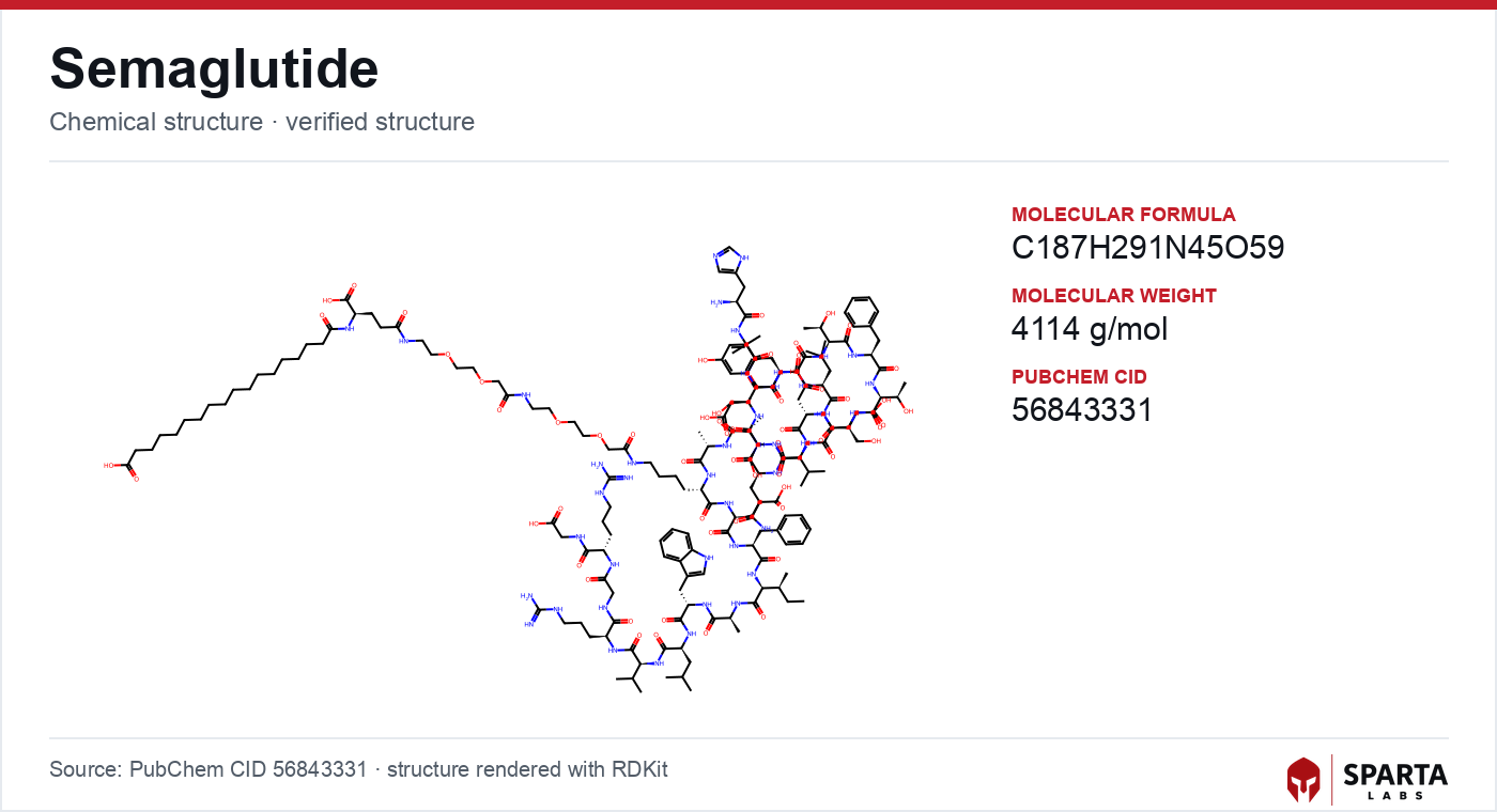
Figure: chemical structure of Semaglutide.
Sequence Engineering: Why the Mechanism Starts with the Molecule
Semaglutide's pharmacology cannot be separated from three specific modifications to the GLP-1 backbone, each documented in the discovery paper by Lau and colleagues [1].
- Aib at position 8. The native GLP-1 peptide is cleaved at the Ala8-Glu9 bond by dipeptidyl peptidase-4 (DPP-4), which is why endogenous GLP-1 has a circulating half-life of roughly one to two minutes. Substituting alanine-8 with the non-proteinogenic residue 2-aminoisobutyric acid (Aib) sterically shields this cleavage site and confers DPP-4 resistance [1].
- C-18 diacid acylation at Lys26. A gamma-glutamate spacer and a 1,2-distearoyl-sn-glycero-derived C-18 fatty diacid are attached through a short linker at lysine-26. This lipid moiety mediates reversible, high-affinity binding to circulating serum albumin [1].
- Arg34 substitution. Replacing lysine-34 with arginine directs the acylation exclusively to Lys26 and removes a competing conjugation site [1].
The net effect is a receptor agonist whose extended residence time in plasma arises from albumin sequestration and protease resistance rather than from any change to the intrinsic on-rate at the receptor. Lau and colleagues reported a GLP-1R binding affinity of approximately 0.38 nM for semaglutide, alongside markedly increased albumin affinity relative to the earlier analogue liraglutide [1].
Findings from research models do not establish safety or efficacy in humans. Sparta Labs makes no claims about the use of this compound.
Engaging the Receptor: The Two-Domain Binding Model
Class B GPCRs such as GLP-1R are distinguished by a large extracellular domain (ECD) that works in concert with the seven-transmembrane core. Structural and biochemical work is generally consistent with a two-domain, or "two-step," binding model: the C-terminal helix of the peptide first docks into the ECD, positioning the N-terminus to insert into the transmembrane bundle, where it triggers the conformational rearrangement that opens the intracellular face to G-protein coupling [2].
Cryo-electron microscopy of the active, peptide-bound GLP-1R in complex with heterotrimeric Gs, reported by Zhang and colleagues in 2017, resolved this arrangement at near-atomic detail and clarified how N-terminal peptide insertion is coupled to outward movement of transmembrane helix 6 [3]. Because semaglutide retains the native GLP-1 N-terminal residues that make these transmembrane contacts, it engages the same orthosteric mechanism as the endogenous ligand; the engineered modifications sit on the peripheral, albumin-facing surface rather than at the receptor interface [1][3].
From cAMP to Effector Arms
Once the active receptor conformation is stabilized, GLP-1R couples predominantly to the stimulatory G-protein Gs, activating adenylyl cyclase and elevating intracellular cyclic adenosine monophosphate (cAMP) as the principal second messenger [4][5]. Drucker's 2018 review in Cell Metabolism organizes the downstream biology into distinguishable effector arms whose relative weighting is tissue-dependent [4].
The cAMP/PKA arm
Elevated cAMP activates protein kinase A (PKA). In pancreatic beta-cells, PKA-mediated phosphorylation events have been reported to contribute to the potentiation of glucose-stimulated insulin secretion [4]. Among the nuclear targets is the cAMP response element-binding protein (CREB); Drucker noted that CREB activation drives expression of genes including brain-derived neurotrophic factor and Bcl-2 in experimental islet models [4].
The EPAC arm
The exchange protein directly activated by cAMP (EPAC2) operates as a PKA-independent effector of GLP-1R-generated cAMP. Pharmacological dissection studies reviewed by Drucker attribute a component of calcium-dependent insulin exocytosis to EPAC2 rather than PKA, indicating parallel rather than strictly linear signaling [4].
Receptor internalization and biased signaling
Beyond G-protein coupling, ligand-bound GLP-1R recruits beta-arrestins, which govern receptor internalization and can scaffold arrestin-dependent signaling. The extent to which different GLP-1R agonists favor sustained cAMP output versus internalization is an active question in the biased-agonism literature and is thought to influence the duration of the signaling response [4][6]. Comparative receptor-signaling profiles across the incretin class are discussed in the tirzepatide mechanism of action and retatrutide mechanism of action articles, which cover dual and triple incretin agonists respectively.
Tissue-Level Consequences Reported in the Literature
Glucose-dependent islet effects
A defining pharmacological feature of GLP-1R agonism is glucose dependence: the potentiation of insulin secretion is substantially attenuated at low plasma glucose, because membrane depolarization and calcium entry are required as permissive signals for exocytosis [4]. Research reviewed by Drucker also reported suppression of glucagon secretion from pancreatic alpha-cells under GLP-1R agonism, though whether this is direct, given that only a subset of alpha-cells express the receptor, or indirect via somatostatin and paracrine signaling, remains under investigation [4].
Gastric motility
GLP-1Rs are expressed on enteric and vagal neurons, and receptor activation has been associated with a slowing of gastric emptying in pharmacological studies [4][6]. This alters postprandial nutrient-absorption kinetics and is mechanistically distinct from the direct islet effects. The relative contribution of gastric-motility effects to the overall pharmacological profile in humans remains an open research question [6].
Central nervous system expression
GLP-1Rs are present in defined hypothalamic and hindbrain nuclei, including the arcuate nucleus and the nucleus of the solitary tract [4][6]. Preclinical work summarized by Nauck and colleagues has implicated these central receptor populations in the integration of energy-balance signals, using genetic and lesion approaches to distinguish central from peripheral receptor contributions [6]. These findings derive predominantly from animal models, and their translation to human neurobiology is an ongoing area of study.
Cardiovascular signaling
GLP-1R expression has been reported in cardiac tissue and vascular endothelium, generally at lower levels than in pancreatic islets [4]. The mechanistic basis for the reduction in major adverse cardiovascular events reported in the SELECT trial has not been fully resolved; proposed contributors discussed in the review literature include anti-inflammatory signaling, effects on endothelial function, and indirect metabolic changes, and each remains an active research area [6][7].
Limits of Current Understanding
Several mechanistic questions define the current frontier. The proportional importance of the Gs/PKA, EPAC, and beta-arrestin arms across different human tissue compartments is not fully resolved, and emerging single-cell and spatial methods are progressively refining GLP-1R expression maps [4][6]. The receptor-trafficking dynamics that govern signaling duration under sustained agonist exposure, and how albumin-mediated pharmacokinetics interact with those dynamics, are areas of continuing inquiry [1][6]. For investigators studying GLP-1R pharmacology, semaglutide from Sparta Labs is offered as a research-grade reference material; structural and analytical verification practices for this compound are described in the semaglutide sourcing and quality article.
References
-
Lau J, Bloch P, Schäffer L, et al. Discovery of the once-weekly glucagon-like peptide-1 (GLP-1) analogue semaglutide. J Med Chem. 2015;58(18):7370–7380. doi:10.1021/acs.jmedchem.5b00726. PubMed PMID: 26308095. PubMed
-
Willard FS, Sloop KW. Physiology and emerging biochemistry of the glucagon-like peptide-1 receptor. Exp Diabetes Res. 2012;2012:470851. doi:10.1155/2012/470851. PubMed PMID: 22577360. PubMed
-
Zhang Y, Sun B, Feng D, et al. Cryo-EM structure of the activated GLP-1 receptor in complex with a G protein. Nature. 2017;546(7657):248–253. doi:10.1038/nature22394. PubMed PMID: 28538729. PubMed
-
Drucker DJ. Mechanisms of action and therapeutic application of glucagon-like peptide-1. Cell Metab. 2018;27(4):740–756. doi:10.1016/j.cmet.2018.03.001. PubMed PMID: 29617641. PubMed
-
Knudsen LB, Lau J. The discovery and development of liraglutide and semaglutide. Front Endocrinol (Lausanne). 2019;10:155. doi:10.3389/fendo.2019.00155. PubMed PMID: 31031702. PubMed
-
Nauck MA, Quast DR, Wefers J, Meier JJ. GLP-1 receptor agonists in the treatment of type 2 diabetes — state-of-the-art. Mol Metab. 2021;46:101102. doi:10.1016/j.molmet.2020.101102. PubMed PMID: 33068776. PubMed
-
Lincoff AM, Brown-Frandsen K, Colhoun HM, et al. Semaglutide and cardiovascular outcomes in obesity without diabetes. N Engl J Med. 2023;389(24):2221–2232. doi:10.1056/NEJMoa2307563. PubMed PMID: 37952131. PubMed
Disclaimer. Statements in this article have not been evaluated by the Food and Drug Administration. This compound is not intended to diagnose, treat, cure, or prevent any disease. Sparta Labs sells research-use-only materials. Content is provided for educational and informational purposes only and does not constitute medical advice. Consult a qualified medical professional for any health concerns.
Frequently asked questions
How does semaglutide's molecular design affect its mechanism?
Three engineered features distinguish semaglutide from native GLP-1: a 2-aminoisobutyric acid (Aib) substitution at position 8 that resists DPP-4 cleavage, a C-18 fatty diacid acylation at Lys26 that binds serum albumin, and an Arg34 substitution that directs acylation to a single site. Lau and colleagues reported that these modifications extend circulating residence time without altering the intrinsic receptor-binding mechanism.
What receptor does semaglutide bind, and how?
Semaglutide binds the glucagon-like peptide-1 receptor (GLP-1R), a class B secretin-family GPCR. Binding follows a two-domain model in which the peptide's C-terminal helix docks into the extracellular domain and its N-terminus inserts into the transmembrane bundle. A 2017 cryo-EM structure by Zhang and colleagues resolved this active, G-protein-coupled arrangement.
What signaling pathways does GLP-1 receptor activation engage?
GLP-1R couples predominantly to the stimulatory G-protein Gs, activating adenylyl cyclase and elevating cyclic AMP. Reviewed literature describes distinguishable effector arms downstream of cAMP, including protein kinase A (PKA), the exchange protein EPAC2, and beta-arrestin-dependent pathways, with tissue-dependent weighting.
Why is the insulin-secretory effect of semaglutide described as glucose-dependent?
GLP-1R agonism potentiates insulin secretion only when membrane depolarization and calcium entry provide permissive signals, which occur at elevated glucose. As Drucker's review notes, the effect is substantially attenuated at low plasma glucose because those permissive conditions are absent.