
Hexarelin Mechanism of Action
A mechanistic reference on hexarelin's unusual two-receptor pharmacology: GHS-R1a-driven calcium and MAPK signaling versus CD36-mediated PPARgamma transcription, and where the two cascades converge on cell-survival endpoints in preclinical research.

For research use only. Not for human consumption. This article is educational reference material. It is not medical advice and is not a recommendation to use any substance.
Why Hexarelin Has Two Receptors
Most growth hormone secretagogues are described in the literature through a single receptor: the ghrelin/growth hormone secretagogue receptor type 1a (GHS-R1a). Hexarelin (also called examorelin) is unusual in that peer-reviewed work has characterized it at a second, structurally unrelated protein, the class B scavenger receptor CD36. This dual-receptor pharmacology is the organizing theme of hexarelin's mechanistic literature, because several reported effects segregate cleanly by receptor: neuroendocrine signaling maps onto GHS-R1a, while a set of peripheral cardiovascular and metabolic observations has been attributed to CD36.
The sections below trace hexarelin from its primary sequence, through each receptor's binding pharmacology, into the divergent intracellular cascades each engages, and finally to the downstream cellular endpoints reported in preclinical models. Where the two pathways converge, the literature is noted as convergent rather than assigned to one receptor. A broader treatment of the compound's classification and discovery is available in the hexarelin research overview.
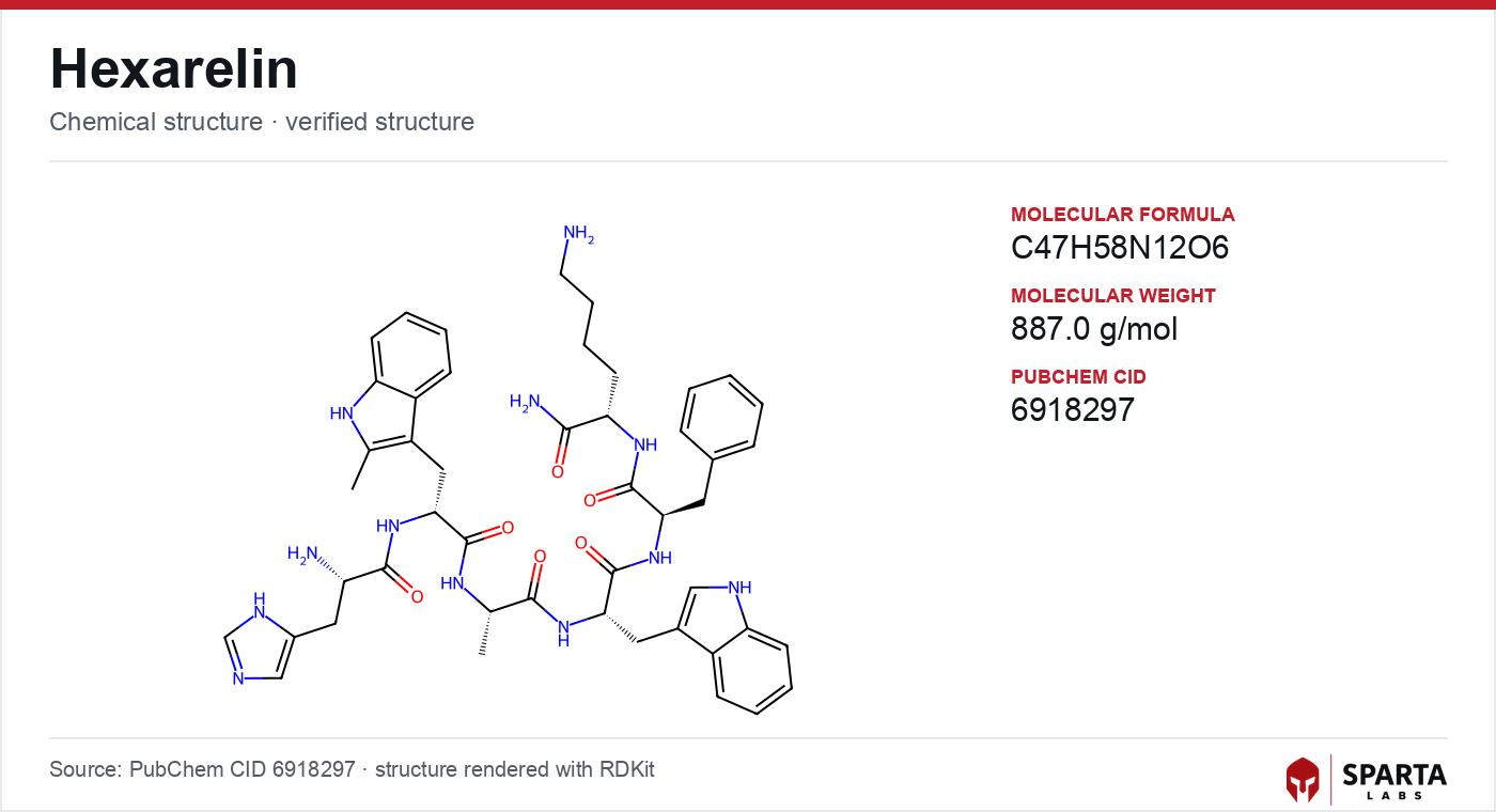
Figure: chemical structure of Hexarelin.
Sequence Design and What It Buys
Hexarelin is a synthetic hexapeptide, His-D-2-methyl-Trp-Ala-Trp-D-Phe-Lys-NH2, developed as an analog of the earlier secretagogue GHRP-6. The single design change most relevant to its mechanism is the substitution at position two: an unnatural D-2-methyltryptophan residue replaces the D-tryptophan of GHRP-6. Deghenghi and colleagues reported in 1994 that this modification was associated with growth-hormone-releasing activity in both infant and adult rat preparations, and the methylation is generally understood to confer resistance to peptidase cleavage relative to the parent peptide.[2]
That stability is mechanistically consequential. A secretagogue's observed potency in an intact model reflects not only receptor affinity but how long the ligand survives to occupy the receptor. The structural relationship between hexarelin and its precursor is discussed further in the GHRP-6 mechanism of action article, and the shared pharmacophore is common to the wider GHRP family, including the compound examined in the GHRP-2 mechanism of action reference.
The GHS-R1a Arm
A receptor found before its ligand
GHS-R1a is a seven-transmembrane G protein-coupled receptor (GPCR) in the rhodopsin family. It was cloned in 1996 by Howard and colleagues, who identified it in pituitary and hypothalamic tissue as the molecular site responsible for the GH-releasing action of synthetic secretagogues, several years before ghrelin was recognized as its endogenous ligand in 1999.[1]
Findings from research models do not establish safety or efficacy in humans. Sparta Labs makes no claims about the use of this compound.
This ordering matters for interpreting hexarelin: the receptor was defined pharmacologically, by the synthetic peptides that activated it, and hexarelin is characterized in the literature as a high-affinity full agonist at this site. GHS-R1a couples predominantly to the Gq/11 family of heterotrimeric G proteins, which is the entry point to its principal signaling cascade.
From receptor occupancy to calcium
Activation of Gq/11 stimulates phospholipase C (PLC), which hydrolyzes the membrane lipid phosphatidylinositol 4,5-bisphosphate (PIP2) into two second messengers: inositol 1,4,5-trisphosphate (IP3) and diacylglycerol (DAG). IP3 mobilizes calcium (Ca2+) from intracellular endoplasmic reticulum stores, while DAG activates protein kinase C (PKC). PKC in turn gates voltage-operated L-type calcium channels, allowing further calcium entry from outside the cell. In anterior pituitary somatotrophs, this rise in intracellular calcium is the proximal trigger for the exocytosis of stored growth hormone.
A kinase branch beyond the pituitary
GHS-R1a signaling is not confined to the calcium/PKC axis. Mousseaux and colleagues reported that receptor activation engaged extracellular signal-regulated kinase 1/2 (ERK1/2) through a PLC/PKCepsilon branch, placing hexarelin's target within the mitogen-activated protein kinase (MAPK) network characteristic of many GPCRs.[5] A separate line of work in Neuro-2A neuronal cells reported that hexarelin modulated both MAPK and the phosphoinositide 3-kinase (PI3K)/Akt pathway, with observed attenuation of hydrogen-peroxide-induced apoptotic toxicity in that model.[6] These kinase observations extend the mechanistic picture past the neuroendocrine setting and into cell-survival signaling.
The CD36 Arm
A second binding site identified by cross-linking
In 2004, Bodart and colleagues reported a photoaffinity cross-linking study that identified CD36 as a distinct hexarelin binding site.[4] CD36, also called fatty acid translocase, is a class B scavenger receptor expressed on cardiomyocytes, monocytes and macrophages, platelets, and microvascular endothelium. In the same assay system, hexarelin and related GHRPs bound CD36 with affinities comparable to their GHS-R1a binding, whereas ghrelin itself showed substantially lower CD36 affinity.
That contrast is the pharmacological crux of the two-receptor model. Because the endogenous GHS-R1a ligand does not engage CD36 appreciably, effects that track with CD36 occupancy cannot be assumed to reflect ordinary ghrelinergic signaling, and hexarelin becomes a tool for probing CD36 biology that ghrelin cannot replicate.
Transcriptional signaling through PPARgamma
CD36 engagement routes into a transcriptional program rather than the acute calcium flux of the GHS-R1a arm. Marleau and colleagues reviewed evidence that hexarelin and related peptides, acting through CD36, triggered activation of the nuclear receptor peroxisome proliferator-activated receptor gamma (PPARgamma), with downstream induction of apolipoprotein E and the sterol transporters ABCA1 and ABCG1, and associated effects on cholesterol efflux in macrophage models.[7] In hepatocyte models, the same review described engagement of the LKB1-AMPK pathway, linking CD36 occupancy to enzymes of cholesterol synthesis. This is a slower, gene-expression-level mode of action that has no counterpart in the pituitary GHS-R1a cascade.
Where the Two Arms Converge: Cell-Survival Signaling
Several preclinical reports describe hexarelin-associated outcomes that converge on cell-survival machinery regardless of which receptor is nominally engaged. In cardiomyocyte models of ischemia-reperfusion, hexarelin treatment was associated with modulation of apoptotic markers, including caspase activity and altered expression of Bcl-2 family proteins.[3] In the Neuro-2A neuronal model noted above, PI3K/Akt activation and MAPK modulation accompanied reduced apoptotic toxicity.[6]
The convergence is analytically important. Because both GHS-R1a (via ERK1/2 and PI3K/Akt) and CD36 (via PPARgamma and AMPK) can feed anti-apoptotic signaling, a survival phenotype observed in an intact cell cannot by itself be attributed to a single receptor without selective pharmacological tools. The literature generally treats these endpoints as convergent rather than receptor-specific.
Downstream Effects Reported in Preclinical Models
Cardiac endpoints and the GH-independence question
A 2014 review in Endocrinology catalogued preclinical cardiovascular observations across multiple experimental systems.[3] Reported outcomes in rodent models included attenuation of post-ischemic ventricular dysfunction, reduced collagen deposition after myocardial injury, modulation of inflammatory cytokine signaling, and preserved cardiomyocyte viability. A recurring feature of this literature is that such effects were reported in preparations where GH secretion was absent or GHS-R1a signaling was pharmacologically altered, which is what motivated the search for a GH-independent pathway. The identification of CD36 in cardiac tissue supplied the mechanistic candidate for those GH-independent observations.
Regulation of the receptor itself
At the hypothalamic-pituitary level, hexarelin's GHS-R1a agonism has been reported to modulate GHS-R1a mRNA expression at both sites in a time-dependent manner following repeated administration.[8] Receptor-level regulation of this kind is consistent with the desensitization and expression dynamics characterized broadly across GPCR pharmacology, and it offers a mechanistic frame for the altered GH-releasing response observed after prolonged exposure in animal studies.
Limits of Current Understanding
Two questions dominate the open literature. The first is attribution: the relative contribution of GHS-R1a versus CD36 to any specific downstream effect remains unresolved in most studies, largely because receptor-selective antagonists and knockout models have not been applied uniformly across research groups. The second is scope: the CD36 signaling network engaged by hexarelin is less thoroughly mapped than the GHS-R1a cascade, and whether the PPARgamma/ABCA1 program described in macrophages operates in cardiomyocyte or endothelial contexts is an experimental question that remains active.
All of the mechanistic work summarized here derives from in vitro systems or rodent in vivo models; extension to human biology is the direction of ongoing clinical interest rather than an established fact. The body of primary studies applying these frameworks is compiled in the hexarelin published research summary, and the timeline from the compound's synthesis through subsequent investigation is covered in the hexarelin discovery and regulatory history. For researchers requiring material of verified identity, hexarelin from Sparta Labs is third-party tested for purity and molecular identity.
References
-
Howard AD, Feighner SD, Cully DF, Arena JP, Liberator PA, Rosenblum CI, et al. A receptor in pituitary and hypothalamus that functions in growth hormone release. Science. 1996;273(5277):974–977. PMID: 8688086. DOI: 10.1126/science.273.5277.974
-
Deghenghi R, Cananzi MM, Torsello A, Battisti C, Müller EE, Locatelli V. GH-releasing activity of Hexarelin, a new growth hormone releasing peptide, in infant and adult rats. Life Sci. 1994;54(18):1321–1328. PMID: 7910650. DOI: 10.1016/0024-3205(94)00845-X
-
Cao Y, Liu L, Fang W, Zhu X, Shi H. The cardiovascular action of hexarelin. Endocrinology. 2014;155(12):4534–4539. PMID: 25278975. DOI: 10.1210/en.2014-1285
-
Bodart V, Bouchard JF, McNicoll N, Escher E, Carrière P, Ghigo E, et al. Identification of the growth hormone-releasing peptide binding site in CD36: a photoaffinity cross-linking study. Biochemistry. 2004;43(18):5557–5565. PMID: 15176951. DOI: 10.1021/bi0302085
-
Mousseaux D, Le Gallic L, Ryan J, Oiry C, Gagne D, Fehrentz JA, et al. Regulation of ERK1/2 activity by ghrelin-activated growth hormone secretagogue receptor 1A involves a PLC/PKCepsilon pathway. Br J Pharmacol. 2006;148(3):350–365. PMID: 16604093. DOI: 10.1038/sj.bjp.0706727
-
Mosa RMH, Zhang Z, Shao R, Deng C, Chen J, Chen C. Hexarelin modulates MAPK and PI3K/Akt pathways in Neuro-2A cells to inhibit hydrogen-peroxide-induced apoptotic toxicity. Int J Mol Sci. 2021;22(9):4955. PMID: 34066780. DOI: 10.3390/ijms22094955
-
Marleau S, Mulumba M, Lamontagne D, Ong H. Hexarelin signaling to PPARgamma in metabolic diseases. PPAR Res. 2007;2007:87489. DOI: 10.1155/2007/87489
-
Moulin A, Demange L, Bergé G, Gagne D, Ryan J, Mousseaux D, et al. Hexarelin modulates the expression of growth hormone secretagogue receptor type 1a mRNA at hypothalamic and pituitary sites. Neuroendocrinology. 2004;79(6):324–332. PMID: 15361691. DOI: 10.1159/000079843
Disclaimer. Statements in this article have not been evaluated by the Food and Drug Administration. This compound is not intended to diagnose, treat, cure, or prevent any disease. Sparta Labs sells research-use-only materials. Content is provided for educational and informational purposes only and does not constitute medical advice. Consult a qualified medical professional for any health concerns.
Frequently asked questions
Why is hexarelin described as a dual-receptor peptide?
Peer-reviewed work has characterized hexarelin at two structurally unrelated proteins: the GHS-R1a ghrelin receptor and the class B scavenger receptor CD36. This distinguishes it from ghrelin, the endogenous GHS-R1a ligand, which shows substantially lower CD36 affinity in the same assay systems. Several reported effects segregate by receptor, with neuroendocrine signaling mapped to GHS-R1a and certain peripheral cardiovascular observations attributed to CD36.
What signaling does hexarelin trigger through GHS-R1a?
GHS-R1a couples to Gq/11, activating phospholipase C, which generates IP3 and DAG. IP3 mobilizes intracellular calcium while DAG activates protein kinase C, and in pituitary somatotrophs this calcium rise is the proximal trigger for growth hormone exocytosis. Published research also reports engagement of the ERK1/2 MAPK cascade through a PLC/PKCepsilon branch and, in neuronal models, PI3K/Akt signaling.
What is CD36 and how does it relate to hexarelin?
CD36, also called fatty acid translocase, is a scavenger receptor expressed on cardiomyocytes, macrophages, platelets, and endothelium. A 2004 photoaffinity cross-linking study identified it as a distinct hexarelin binding site with affinity comparable to GHS-R1a. Through CD36, hexarelin has been associated with PPARgamma transcriptional activation and, in hepatocyte models, LKB1-AMPK engagement.
Why is the growth-hormone-independent pathway of hexarelin discussed in the literature?
Reviews have catalogued cardiovascular observations in rodent models where GH secretion was absent or GHS-R1a signaling was pharmacologically altered, indicating some effects did not depend on growth hormone release. The identification of CD36 in cardiac tissue provided a candidate mechanism for these GH-independent observations. All such findings derive from preclinical systems and do not establish outcomes in humans.