
Kisspeptin-10: Mechanism of Action
A mechanistic reference on kisspeptin-10 at KISS1R, tracing the RFamide pharmacophore, Gq/11-phospholipase C signaling, TRPC-mediated depolarization, and GnRH neuron firing described across primary neuroendocrine literature. Educational reference.
For research use only. Not for human consumption. This article is educational reference material. It is not medical advice and is not a recommendation to use any substance.
Introduction
Kisspeptin-10 (KP-10) is the C-terminal decapeptide fragment of the larger kisspeptin precursor and a full agonist at the kisspeptin receptor KISS1R, a class A (rhodopsin-family) G-protein-coupled receptor. Within reproductive neuroendocrinology, KP-10 has become a widely used pharmacological probe because it reproduces the receptor-activating activity of the full-length peptide while being short enough for straightforward synthesis. This article traces the reported molecular mechanism of KP-10 in a stepwise fashion: from the structural features that let a ten-residue fragment engage its receptor, through the Gq/11-linked second-messenger cascade, to the ion-channel events that alter the firing of gonadotropin-releasing hormone (GnRH) neurons. Each step is attributed to primary peer-reviewed literature. For chemistry, classification, and regulatory context, the companion Kisspeptin-10 research overview provides background beyond the scope of this mechanistic account.
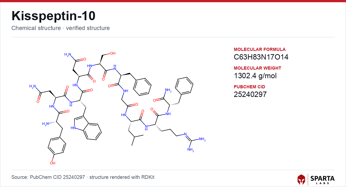
Figure: chemical structure of Kisspeptin-10.
The RFamide Pharmacophore and Receptor Identity
KISS1R (Gene ID: KISS1R; formerly the orphan receptor GPR54) is the receptor for kisspeptin peptides. Kotani and colleagues reported in 2001 that the KiSS-1 gene product is proteolytically processed into kisspeptins that act as the natural ligands of GPR54, establishing the ligand-receptor pairing that underlies all subsequent mechanistic work [1].
The molecular reason a ten-residue fragment is sufficient lies in where the binding determinants reside. The receptor-engaging pharmacophore is concentrated in the shared C-terminal decapeptide, so KP-10 and the longer isoforms (KP-54, KP-14, KP-13) exhibit broadly comparable receptor activity in reported binding studies [1]. The C-terminal arginine-phenylalanine amide (Arg-Phe-NH₂, "RFamide") motif places kisspeptins within the wider RFamide neuropeptide superfamily, and this motif is a recurring structural determinant for receptor engagement across that family.
Findings from research models do not establish safety or efficacy in humans. Sparta Labs makes no claims about the use of this compound.
Anatomically, the mechanistic relevance of KISS1R depends on where the receptor is expressed. Immunohistochemical and in situ hybridization work documented KISS1R transcript alongside GnRH neurons in the mouse hypothalamus, providing the anatomical substrate for a direct action of kisspeptin on these cells rather than a purely indirect, multi-synapse effect [2].
The Core Cascade: Gq/11 and Phospholipase C
Once KP-10 occupies KISS1R, the receptor couples preferentially to the heterotrimeric G-protein Gq/11 and activates phospholipase C beta (PLCβ). PLCβ hydrolyzes the membrane phospholipid phosphatidylinositol 4,5-bisphosphate (PIP₂) into two second messengers: inositol 1,4,5-trisphosphate (IP₃) and diacylglycerol (DAG). Colledge reviewed this canonical Gq cascade in 2009 as the best-characterized signaling axis downstream of the receptor [3], and Pinilla and colleagues later placed it within a comprehensive review of kisspeptin physiology and regulatory mechanisms [4].
The two second messengers diverge functionally. IP₃ binds IP₃ receptors on the endoplasmic reticulum, releasing calcium (Ca²⁺) from intracellular stores. DAG activates protein kinase C (PKC), which phosphorylates downstream effectors including the mitogen-activated protein kinases ERK1/2 and p38, a coupling reported in mechanistic studies of the receptor [4]. This calcium-plus-kinase output is the biochemical bridge between receptor occupancy and the changes in membrane excitability described below.
Translating Signaling into GnRH Neuron Firing
The distinctive feature of KP-10 mechanism in GnRH neurons is how the Gq/11-PLCβ cascade reshapes ion-channel behavior to produce prolonged excitation. Pielecka-Fortuna and Moenter reported in 2012 that kisspeptin activates transient receptor potential canonical (TRPC) channels in female GnRH neurons through a mechanism requiring PIP₂ depletion and cSrc tyrosine-kinase activation [5]. Opening these non-selective cation channels admits an inward, depolarizing current.
The same line of work reported that KP-10 concurrently suppresses potassium (K⁺) currents, including A-type and inwardly rectifying components, reducing the outward current that would otherwise repolarize the cell [5]. Xu and colleagues added resolution in 2018, describing a phospholipase C and calcium-dependent pathway that regulates multiple ion channels in concert to drive excitation [6]. The reported net effect of inward TRPC current combined with reduced K⁺ conductance is a sustained depolarization and an elevated action-potential firing rate in GnRH neurons. This coordination of several channel families by a single receptor pathway distinguishes the kisspeptin response from a simple, transient synaptic potential.
Where the Signal Is Delivered: Soma Versus Nerve Terminal
Kisspeptin does not act only at the GnRH neuron cell body. Irwig and colleagues demonstrated in 2004 that kisspeptin stimulates GnRH release directly at nerve terminals in the male rat, using a preparation of isolated neurosecretory terminals to isolate a presynaptic locus of action [7]. Identifying a terminal-level site of action at the median eminence, in addition to the soma, refined the circuit model of how the peptide couples to hormone output.
This distinction carries through to route-dependent observations. Studies comparing administration routes in rodents reported that central delivery elicits c-Fos expression in GnRH neurons whereas peripheral delivery does not, an asymmetry interpreted as consistent with kisspeptin acting at GnRH terminals in the median eminence rather than at the soma [2]. Because GnRH released into the hypophysial portal circulation drives luteinizing hormone (LH) and follicle-stimulating hormone secretion, plasma LH is commonly used as an indirect functional readout of GnRH neuron activation in these research designs.
KNDy Circuitry and the Pulse-Generator Model
KP-10 mechanism is often studied in the context of the KNDy neuron population, arcuate-nucleus cells that co-express kisspeptin, neurokinin B, and dynorphin A. Lehman and colleagues reviewed in 2010 a model in which KNDy neurons function as a pulse generator, with kisspeptin acting as the output signal to GnRH neurons [8]. Under this framework, exogenous KP-10 serves as a tool to acutely activate KISS1R at the GnRH output stage and probe the timing properties of the hypothalamic-pituitary-gonadal axis. The experimental evidence bearing on this model is surveyed in the Kisspeptin-10 published-research summary, and the historical development of the KNDy concept is traced in the discovery and research history article.
Non-Canonical Coupling and Open Kinetic Questions
While the Gq/11-PLCβ axis is the best-supported signaling route, reviews note reports of additional coupling to other G-protein families in heterologous expression systems, raising the possibility of context-dependent signaling diversity in native GnRH neurons [4]. Whether such alternative coupling operates in situ remains an active question. Comparable context-dependent coupling has been examined in other hypothalamic neuropeptide systems, including the oxytocin mechanism of action, offering a useful point of comparison for GPCR signaling behavior.
Kinetics form a second frontier. Liu and colleagues reported in 2013 an estimated plasma half-life of approximately four minutes for KP-10 in rats, reflecting rapid enzymatic degradation of the decapeptide [9]. This short persistence has motivated research into modified analogs with extended in vivo activity and complicates the interpretation of sustained-infusion versus bolus experiments, since receptor desensitization and internalization dynamics interact with ligand availability. Researchers evaluating peptide identity and integrity for such studies may consult the Kisspeptin-10 sourcing and verification standards reference; the corresponding catalog listing is available at Kisspeptin-10.
References
-
Kotani M, Detheux M, Vandenbogaerde A, Communi D, Vanderwinden JM, Le Poul E, et al. The metastasis suppressor gene KiSS-1 encodes kisspeptins, the natural ligands of the orphan G protein-coupled receptor GPR54. J Biol Chem. 2001;276(37):34631-34636. PMID: 11457843. DOI: 10.1074/jbc.M104847200. PubMed
-
Smith JT, Cunningham MJ, Rissman EF, Clifton DK, Steiner RA. Regulation of Kiss1 gene expression in the brain of the female mouse. Endocrinology. 2005;146(9):3686-3692. PMID: 15919743. DOI: 10.1210/en.2005-0488. PubMed
-
Colledge WH. Kisspeptins and GnRH neuronal signalling. Trends Endocrinol Metab. 2009;20(3):115-121. PMID: 19272794. DOI: 10.1016/j.tem.2008.10.005. PubMed
-
Pinilla L, Aguilar E, Dieguez C, Millar RP, Tena-Sempere M. Kisspeptins and reproduction: physiological roles and regulatory mechanisms. Physiol Rev. 2012;92(3):1235-1316. PMID: 22811428. DOI: 10.1152/physrev.00037.2010. PubMed
-
Pielecka-Fortuna J, Moenter SM. Kisspeptin activation of TRPC4 channels in female GnRH neurons requires PIP2 depletion and cSrc kinase activation. Endocrinology. 2012;153(6):2577-2588. PMID: 22518060. DOI: 10.1210/en.2011-1905. PubMed
-
Xu C, Roepke TA, Zhang C, Kelly MJ, Rønnekleiv OK. Kisspeptin excites gonadotropin-releasing hormone (GnRH) neurons through a phospholipase C/calcium-dependent pathway regulating multiple ion channels. Endocrinology. 2018;159(9):3265-3279. PMID: 30016398. DOI: 10.1210/en.2018-00315. PubMed
-
Irwig MS, Fraley GS, Smith JT, Acohido BV, Popa SM, Cunningham MJ, et al. Kisspeptin activation of gonadotropin releasing hormone neurons and regulation of KiSS-1 mRNA in the male rat. Neuroendocrinology. 2004;80(4):264-272. PMID: 15665093. DOI: 10.1159/000083140. PubMed
-
Lehman MN, Coolen LM, Goodman RL. Minireview: kisspeptin/neurokinin B/dynorphin (KNDy) cells of the arcuate nucleus: a central node in the control of gonadotropin-releasing hormone secretion. Endocrinology. 2010;151(8):3479-3489. PMID: 20501670. DOI: 10.1210/en.2010-0022. PubMed
-
Liu Z, Ren C, Jones W, Chen P, Seminara SB, Chan YM, et al. LC-MS/MS quantification of a neuropeptide fragment kisspeptin-10 (NSC 741805) and characterization of its decomposition product and pharmacokinetics in rats. J Chromatogr B Analyt Technol Biomed Life Sci. 2013;926:1-8. PMID: 23524040. DOI: 10.1016/j.jchromb.2013.02.027. PubMed
Disclaimer. Statements in this article have not been evaluated by the Food and Drug Administration. This compound is not intended to diagnose, treat, cure, or prevent any disease. Sparta Labs sells research-use-only materials. Content is provided for educational and informational purposes only and does not constitute medical advice. Consult a qualified medical professional for any health concerns.
Frequently asked questions
What receptor does kisspeptin-10 bind, and why is its C-terminus important?
Published research identifies KISS1R (formerly GPR54) as the receptor for kisspeptin peptides, including kisspeptin-10. Kotani and colleagues reported in 2001 that the C-terminal decapeptide carries the receptor-binding pharmacophore, with the arginine-phenylalanine amide (RFamide) motif characteristic of this neuropeptide family. This is why the truncated 10-residue fragment retains receptor engagement comparable to the longer kisspeptin isoforms.
What intracellular signaling pathway is reported downstream of KISS1R?
Reviews of the primary literature describe KISS1R coupling preferentially to the Gq/11 protein, which activates phospholipase C beta. Phospholipase C beta hydrolyzes PIP2 into IP3 and diacylglycerol, mobilizing intracellular calcium and activating protein kinase C. Colledge summarized this canonical cascade in 2009 as the best-characterized signaling axis for the receptor.
How is kisspeptin-10 reported to change GnRH neuron electrical activity?
Pielecka-Fortuna and Moenter reported in 2012 that kisspeptin-10 gates transient receptor potential canonical (TRPC) channels in GnRH neurons through PIP2 depletion and cSrc kinase activation, while reducing potassium currents. Xu and colleagues in 2018 described a phospholipase C and calcium-dependent pathway coordinating multiple ion channels. The reported net effect in these models is sustained membrane depolarization and increased action potential firing.
Why do researchers study the short plasma half-life of kisspeptin-10?
Liu and colleagues reported in 2013 an estimated plasma half-life of roughly four minutes in rats, reflecting rapid enzymatic degradation of the decapeptide. This kinetic feature is relevant to designing modified analogs with extended in vivo persistence and to interpreting acute versus sustained-exposure experiments on the reproductive neuroendocrine axis.