
Oxytocin (Acetate Salt): Mechanism of Action
A structure-informed account of oxytocin receptor pharmacology, from the cyclic nonapeptide binding pose to Gq/11 and Gi/o signaling divergence, drawn from primary literature. Educational reference.
For research use only. Not for human consumption. This article is educational reference material. It is not medical advice and is not a recommendation to use any substance.
Introduction
Oxytocin is a cyclic nonapeptide hormone whose reported biological actions are mediated by a single characterized target, the oxytocin receptor (OXTR). Unlike compounds whose mechanism was inferred largely from downstream phenotype, OXTR pharmacology sits on an unusually deep foundation: decades of radioligand-binding and second-messenger studies were consolidated by a landmark 2001 review, and in 2022 a near-atomic cryo-electron microscopy structure of the active receptor-ligand complex converted much of that inferential model into direct molecular observation. This article traces the reported mechanism from the peptide's binding pose through its divergent G-protein coupling and second-messenger cascades, using peer-reviewed primary literature. A companion summary of preclinical and clinical study designs appears in the oxytocin published research article, and background chemistry is covered in the oxytocin research overview.
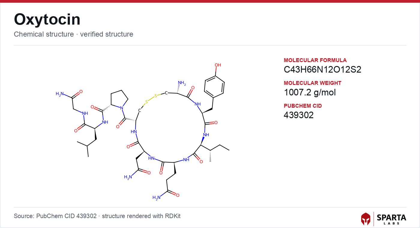
Figure: chemical structure of oxytocin.
The Cyclic Nonapeptide as a Ligand
The pharmacology of oxytocin begins with its constrained geometry. The molecule comprises nine amino acids arranged as a six-residue ring closed by a disulfide bond between the cysteine residues at positions 1 and 6, with a three-residue C-terminal tail extending from the ring. Gimpl and Fahrenholz (2001), in their comprehensive review in Physiological Reviews, summarized early structure-activity work indicating that integrity of the disulfide-bridged ring is required for high-affinity receptor engagement, and that oxytocin differs from the closely related vasopressin peptides by only two residues [1].
Findings from research models do not establish safety or efficacy in humans. Sparta Labs makes no claims about the use of this compound.
That two-residue difference is central to how the field frames receptor selectivity. Oxytocin and the three vasopressin receptor subtypes share substantial sequence homology, and the modest chemical distinction between the native ligands is what pharmacologists have long invoked to explain the cross-reactivity observed at high concentrations [1]. Understanding the binding determinants that discriminate OXTR from the vasopressin receptors has therefore been a recurring objective of structural investigation.
What the Cryo-EM Structure Revealed
For much of its history, the OXTR binding pose was modeled indirectly from mutagenesis and homology to other class A receptors. Waltenspuhl and colleagues (2022) reported single-particle cryo-electron microscopy structures of the active human oxytocin receptor bound to its cognate ligand, resolving the complex at a level that allowed direct assignment of contact residues [2]. The structure indicated that all nine amino acids of oxytocin participate in the interaction, with the cyclic ring (residues 1 through 6) buried within the transmembrane binding cavity and the carboxy-terminal tripeptide (residues 7 through 9) oriented toward the extracellular loop region.
The same study described a magnesium ion coordinated within the ligand-receptor interface, an element that had not been resolved by earlier approaches. The authors discussed this cation-coordination feature in the context of receptor activation and selectivity relative to the vasopressin subtypes [2]. The structure thus reframed selectivity as a question not only of the two divergent residues but of the specific network of contacts and the coordinated ion that stabilize the active state.
Divergent G-Protein Coupling
A defining feature of OXTR is that it does not signal through a single G-protein pathway. Jurek and Neumann (2018), reviewing the receptor from intracellular signaling to behavior in Physiological Reviews, described OXTR as coupling principally to the Gq/11 family in most cellular contexts while also engaging Gi/o proteins depending on cell type and receptor expression density [3]. This dual coupling is the mechanistic root of the functional diversity observed across tissues.
The consequence is that the same ligand can produce qualitatively different intracellular outcomes depending on where the receptor is expressed. In systems where Gq/11 predominates, activation drives a calcium-mobilizing cascade; where Gi/o contributes, the signal instead modulates cyclic nucleotide levels. Jurek and Neumann noted that the relative contribution of each pathway in native tissue in vivo, as distinct from receptor-overexpressing cell lines, remains a subject of active pharmacological study [3].
The Phospholipase C-Calcium Cascade
The canonical Gq/11 branch of OXTR signaling follows the pattern established for that G-protein family. Activation stimulates phospholipase C-beta (PLC-beta), which hydrolyzes membrane phosphatidylinositol 4,5-bisphosphate (PIP2) into two second messengers: inositol 1,4,5-trisphosphate (IP3) and diacylglycerol (DAG) [1,3]. IP3 promotes release of calcium from endoplasmic reticulum stores, raising cytosolic calcium concentration, while DAG activates protein kinase C (PKC).
In uterine myometrium, Gimpl and Fahrenholz (2001) described this calcium-mobilizing cascade as the pharmacological basis for the reported contractile response to oxytocin [1]. The calcium rise has additional consequences beyond contraction: Jurek and Neumann (2018) reported that calcium/calmodulin-dependent protein kinase II (CaMKII) can be engaged downstream of OXTR-mediated calcium release, linking the receptor to transcriptional regulation in neuronal preparations [3].
MAPK Signaling and Cyclic-Nucleotide Modulation
Beyond the immediate calcium branch, OXTR activation has been reported to feed into the mitogen-activated protein kinase (MAPK) system. Jurek and Neumann (2018) described activation of extracellular signal-regulated kinase (ERK1/2) downstream of receptor engagement, along with regulation of transcription factors including CREB and MEF-2 and reported effects on neurite outgrowth in neural cell models [3]. These outputs illustrate how a single receptor can connect to programs of gene expression rather than only to acute ionic responses.
The Gi/o-coupled arm has been examined most prominently in cardiac tissue. Gutkowska and colleagues (2000), in a review characterizing oxytocin as a cardiovascular hormone, summarized reports that OXTR activation in animal cardiac preparations was associated with atrial natriuretic peptide release and with negative chronotropic and inotropic effects, attributed to Gi/o-mediated inhibition of adenylyl cyclase and consequent reduction of cyclic AMP [4]. This cardiac line of work is one of the clearer illustrations of how the receptor's second, non-Gq pathway manifests in a specific tissue.
Selectivity, Regulation, and Open Mechanistic Questions
Several mechanistic questions remain unsettled in the primary literature. Jurek and Neumann (2018) identified the kinetics of OXTR desensitization and internalization, and the possibility of receptor dimerization with other GPCRs including vasopressin receptor subtypes, as directions requiring further study [3]. Cross-talk with the vasopressin system is a persistent theme precisely because the ligands and receptors are so structurally close, a point reinforced by the cation-coordination and contact-residue detail from the 2022 structure [2].
Species differences add a further layer. Reported variation in OXTR binding affinity, G-protein coupling preference, and tissue distribution between rodents, non-human primates, and humans has shaped how investigators design and interpret model systems [1,3]. A related node in the same hypothalamic neuroendocrine circuitry is discussed in the kisspeptin-10 mechanism of action article, which describes KISS1R signaling upstream of gonadotropin regulation. Chemistry, salt form, and analytical characterization for research-grade oxytocin acetate are detailed in the oxytocin sourcing and quality article in this library.
References
-
Gimpl G, Fahrenholz F. The oxytocin receptor system: structure, function, and regulation. Physiol Rev. 2001;81(2):629–683. PMID: 11274341. DOI: 10.1152/physrev.2001.81.2.629
-
Waltenspuhl Y, Ehrenmann J, Vacca S, Thom C, Medalia O, Pluckthun A. Structural basis for the activation and ligand recognition of the human oxytocin receptor. Nat Commun. 2022;13(1):4153. PMID: 35851571. PMCID: PMC9293896. DOI: 10.1038/s41467-022-31325-0
-
Jurek B, Neumann ID. The oxytocin receptor: from intracellular signaling to behavior. Physiol Rev. 2018;98(3):1805–1908. PMID: 29897293. DOI: 10.1152/physrev.00031.2017
-
Gutkowska J, Jankowski M, Mukaddam-Daher S, McCann SM. Oxytocin is a cardiovascular hormone. Braz J Med Biol Res. 2000;33(6):625–633. PMID: 10829090. DOI: 10.1590/s0100-879x2000000600003
Disclaimer. Statements in this article have not been evaluated by the Food and Drug Administration. This compound is not intended to diagnose, treat, cure, or prevent any disease. Sparta Labs sells research-use-only materials. Content is provided for educational and informational purposes only and does not constitute medical advice. Consult a qualified medical professional for any health concerns.
Frequently asked questions
What receptor does oxytocin bind, and how is it classified?
Oxytocin binds the oxytocin receptor (OXTR), a class A (rhodopsin-like) G-protein coupled receptor. The human receptor is a 388-amino acid protein with seven transmembrane helices encoded by a single gene on chromosome 3p25. Gimpl and Fahrenholz (2001) reviewed its architecture and tissue distribution across myometrium, mammary myoepithelium, kidney, heart, and several brain regions.
How does the cyclic structure of oxytocin relate to receptor binding?
Oxytocin is a cyclic nonapeptide closed by a disulfide bridge between cysteine residues 1 and 6. Waltenspuhl and colleagues (2022) reported a cryo-EM structure in which the cyclic ring sits deep in the transmembrane binding pocket while the C-terminal tripeptide faces the extracellular loops. Their data indicated all nine residues contribute to binding, and they described a magnesium coordination site within the complex.
What is the difference between Gq/11 and Gi/o coupling at OXTR?
OXTR couples to more than one G-protein family. Jurek and Neumann (2018) reviewed evidence that Gq/11 coupling activates phospholipase C-beta to raise intracellular calcium, whereas Gi/o coupling modulates adenylyl cyclase and cyclic AMP. The balance between these pathways reported in the literature varies with cell type and receptor density.
Which downstream signaling cascades has OXTR activation been associated with?
Reported downstream events include phospholipase C-beta hydrolysis of PIP2 to generate IP3 and diacylglycerol, IP3-driven calcium release, protein kinase C activation, and MAPK/ERK1/2 signaling. Jurek and Neumann (2018) also described CaMKII activation and regulation of transcription factors such as CREB in neuronal preparations.